

Abstract
Learned MRI reconstruction fills information missing from an accelerated acquisition using statistical structure learned from examples. A visually plausible image can therefore contain features weakly constrained by the measured data. This review connects reconstruction datasets and algorithms with null-space analysis, showing why data consistency, image similarity and clinical fidelity require separate evaluation.
A reconstruction contains measured and inferred information
An accelerated MRI acquisition samples only part of the information needed for a fully determined image. Reconstruction combines those measurements with assumptions about likely images. Learning can make those assumptions powerful, but it does not turn unmeasured components into directly observed anatomy. The distinction matters when a small feature is scientifically or clinically important.
Datasets, methods and instability
The fastMRI dataset described by Knoll and colleagues supports reproducible research in accelerated reconstruction. Hammernik and colleagues developed a variational network for MRI reconstruction. Antun and colleagues demonstrated instabilities in learned image reconstruction under selected perturbations and distribution changes. These contributions motivate evaluation beyond average image-similarity scores. [1], [2], [3]
Data consistency has a null space
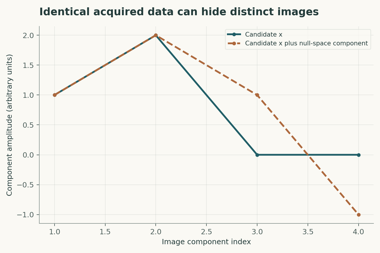
If a perturbation lies in the null space of the acquisition operator, adding it to an image does not change the ideal measured data. Two different images can therefore both be perfectly data consistent. A prior chooses between them. Data consistency is necessary for many reconstruction approaches, but it is not sufficient to prove that every inferred feature is correct.
Global error can ignore a small lesion
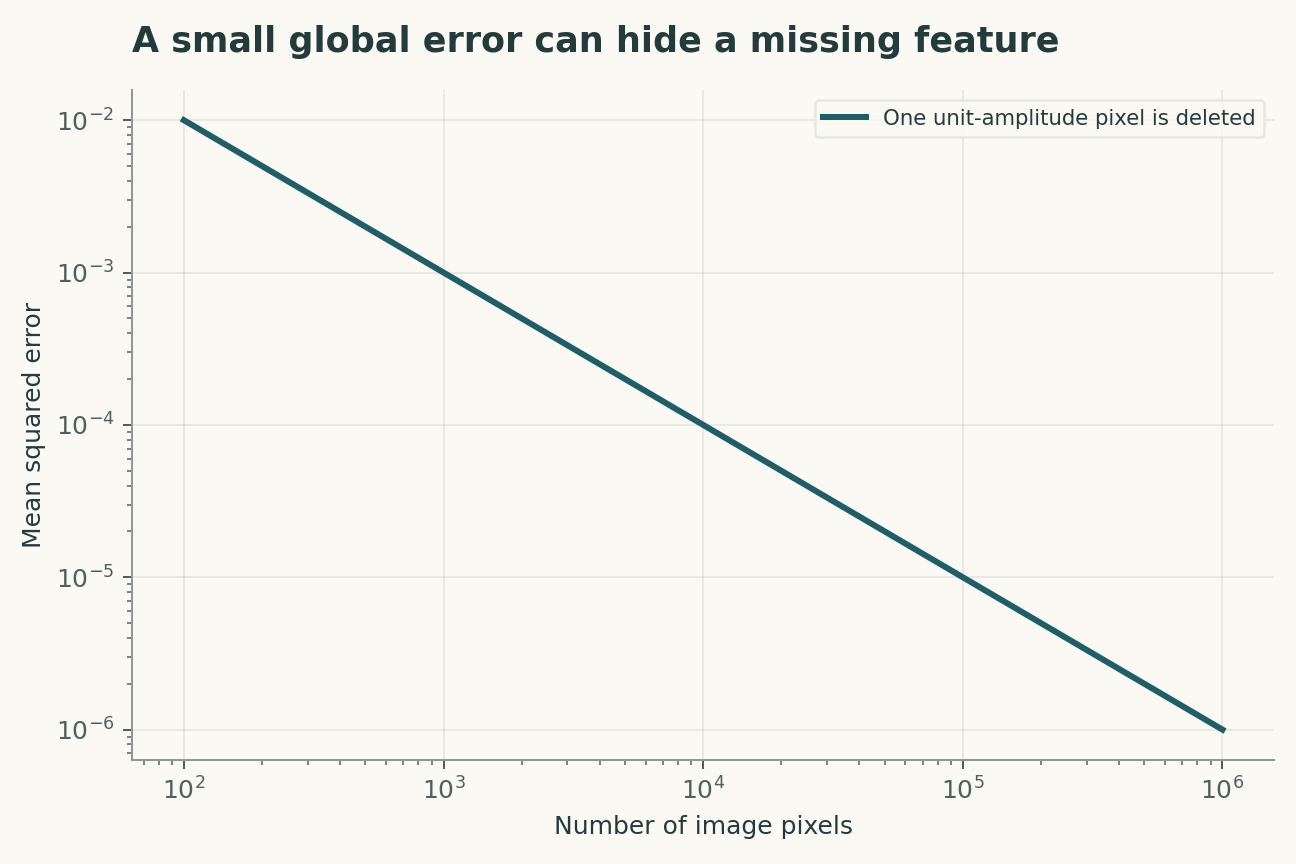
Mean squared error averages over all pixels. A small, important feature can be removed while the global score changes little, especially if smoothing improves a large background region. A task-specific evaluation should test feature detection, localization and uncertainty, not only overall fidelity. The target feature and its acceptable error need to be defined before model selection.
Sampling masks create a distribution
A model trained under one sampling pattern may respond differently to another acceleration factor, coil configuration or noise level. Retrospectively removing data from a fully sampled scan can be useful, but it may not reproduce all effects of a prospective accelerated acquisition. Both forms of testing have roles, and their scopes should be stated.
Perturbation tests need physical grounding
Adversarial examples reveal mathematical vulnerabilities, but deployment evaluation should also include plausible motion, calibration error and hardware variation. A model that survives one perturbation family can still fail another. Perturbations should be propagated through the measurement process, not only added to the final image where their meaning may differ.
Uncertainty must be tested against errors
Multiple reconstructions or a confidence map can communicate ambiguity, but their calibration requires independent examples where error can be assessed. A narrow distribution generated by one model may simply repeat the same prior. Uncertainty should expand for unfamiliar acquisitions and weakly constrained features if it is to support meaningful review.
Clinical fidelity is a separate study
A benchmark improvement does not establish diagnostic safety. Reader studies, prospective testing and subgroup analyses address different questions from algorithmic reconstruction error. This review supplies a mathematical evaluation framework rather than a clinical recommendation or a claim that a particular model is validated for patient care.
Data consistency constrains only the measured subspace
If an acquisition operator has a null space, adding a component in that space does not change the acquired data. A reconstruction prior chooses among those compatible images. This is useful when the prior captures real structure, but dangerous when an unusual lesion lies outside its training distribution. A proposed evaluation would insert controlled features before the acquisition operator and examine their recovery across location, size and contrast. Adding features after reconstruction tests display quality rather than measurement recovery. The four-component example makes the ambiguity exact; a realistic test would use the actual sampling and noise model.
Clinical usefulness needs feature-level error analysis
A low average image error can coexist with a missing small structure, as the one-pixel calculation demonstrates. Conversely, a visually conspicuous artifact may have little effect on a particular quantitative task. A proposed evaluation would specify the downstream measurement or diagnostic task, compare against a suitable reference, and analyze errors by feature class and patient subgroup. Reader studies should be blinded and should preserve the independence of patients rather than count multiple slices as separate cases. The purpose is not to replace global image metrics, but to connect them to the information the image is meant to preserve.
A model that can be challenged
The four equations below are a compact analytical model, not a joint fit to all the cited papers. Their interpretation is as follows. MRI measurements y depend on image x through an acquisition operator A that includes sampling and coil response. A learned regularizer or equivalent prior supplies information beyond the measured samples. Null-space perturbations h are invisible to the ideal acquisition, so exact data consistency does not uniquely identify the image. Global squared error weights pixels equally and may underweight a small task-critical feature.
The observational target is measurement fidelity and preservation of task-critical image features. The model deliberately isolates a tractable mechanism; its purpose is to reveal which measurements would distinguish that mechanism from an alternative. Parameter values in the figures are declared illustrative inputs. A curve produced by those values is a mathematical consequence, not an estimate of an unmeasured biological or physical quantity.
A discriminating validation design
The primary comparison should test feature preservation under physically plausible acquisition perturbations. The unit of independent replication is independent patient acquisition; repeated readings from the same unit improve precision but do not create independent replication. A strong validation would hold out new scanners, masks and pathologies before choosing thresholds or fitting nuisance corrections. In particular, acquisition null space and training-distribution shift must be measured, constrained or varied in a sensitivity analysis. Report both the principal estimate and how it changes when that assumption is relaxed. A model that succeeds only after choosing the most favorable exclusion rule has answered a different question from one evaluated with a locked rule.
Worked numerical analysis
In a four-component example, an operator observing only the first two components cannot distinguish images differing only in the last two. Both have zero residual on measured data. Separately, deleting a unit-amplitude feature occupying one pixel of a 10,000-pixel image adds only 0.0001 to MSE. The figures show measurement equivalence and dilution of local error; no synthetic image is presented as a patient scan.
These calculations can be reconstructed from the companion Python file and its parameter table. Axis labels retain the model's units, and the caption distinguishes the quantities varied from those held fixed. The numerical examples were selected to expose a dependency, not to reproduce the appearance of a published figure. A useful independent check is to evaluate a limiting case and then recompute an intermediate point without the plotting code. Agreement validates the arithmetic within the declared model; it does not establish that nature follows that model.
Evidence, transfer and next steps
The scope of the conclusion is limited by acquisition null space and training-distribution shift. Transfer to new scanners, masks and pathologies requires evidence beyond fitting the original observations. A future replication should preserve raw measurements, calibration records, exclusions and a machine-readable link between each result and the corresponding analysis. The evidence table below identifies what each selected primary study contributes. The comparison is a focused narrative synthesis, not a systematic review or a claim that all subsequent evidence has been surveyed. The proposed study has not been performed here. Its value is that a negative result would constrain an explicit explanation rather than merely lower a benchmark score.
A declared analytical model
Four equations, explicit assumptions
(1) MRI measurements y depend on image x through an acquisition operator A that includes sampling and coil response.
(2) A learned regularizer or equivalent prior supplies information beyond the measured samples.
(3) Null-space perturbations h are invisible to the ideal acquisition, so exact data consistency does not uniquely identify the image.
(4) Global squared error weights pixels equally and may underweight a small task-critical feature.
Constructed examples · Reproducible data
Inspect the quantitative argument
These curves are calculated from the declared models. They are not observations from the cited studies. Every figure includes its numeric data and vector version.


Which evidence answers which question?
| Evidence or design choice | What it identifies | What remains unresolved |
|---|---|---|
| Source 1 | fastMRI provided a dataset for reproducible accelerated-reconstruction research. | Dataset performance does not establish clinical deployment validity. |
| Source 2 | A variational network learned an MRI reconstruction procedure. | Training acquisition and population define the learned distribution. |
| Source 3 | Instability analysis demonstrated failure modes under selected perturbations. | The result motivates broader testing rather than a blanket claim about every model. |
| Primary observable | measurement fidelity and preservation of task-critical image features | acquisition null space and training-distribution shift |
| Independent test | feature preservation under physically plausible acquisition perturbations | Requires new measurements |
| Transfer set | new scanners, masks and pathologies | Not validated by the constructed numerical example |
A proposed validation protocol
This is a proposed evaluation design. No new biological experiment, clinical trial or physical experiment was carried out for this article.
- Specify measurement fidelity and preservation of task-critical image features before fitting.
- Retain independent identifiers for each independent patient acquisition.
- Separate new scanners, masks and pathologies from model development.
- Measure or bound acquisition null space and training-distribution shift.
- Record units, calibration and the observation operator.
- Compare the primary model with a named rival explanation.
- Prespecify the test: feature preservation under physically plausible acquisition perturbations.
- Archive raw data, exclusions, parameter files and executable analysis.
What the primary sources establish
Florian Knoll, Jure Zbontar, Anuroop Sriram et al. · 2020 [1]
Contribution. fastMRI provided a dataset for reproducible accelerated-reconstruction research.
Boundary. Dataset performance does not establish clinical deployment validity.
Source locator. Abstract / selected primary-source sections; see linked bibliographic record.
Kerstin Hammernik, Teresa Klatzer, Erich Kobler et al. · 2017 [2]
Contribution. A variational network learned an MRI reconstruction procedure.
Boundary. Training acquisition and population define the learned distribution.
Source locator. Abstract / selected primary-source sections; see linked bibliographic record.
Vegard Antun, Francesco Renna, Clarice Poon et al. · 2020 [3]
Contribution. Instability analysis demonstrated failure modes under selected perturbations.
Boundary. The result motivates broader testing rather than a blanket claim about every model.
Source locator. Abstract / selected primary-source sections; see linked bibliographic record.
Limits of this analysis
- Dataset performance does not establish clinical deployment validity.
- Training acquisition and population define the learned distribution.
- The result motivates broader testing rather than a blanket claim about every model.
- Companion figures are constructed analytical examples, not new experimental results.
- Transfer to new scanners, masks and pathologies has not been demonstrated by this review.
A focused narrative analysis of the named primary studies, not a systematic literature search or a claim to comprehensive coverage through 2026.
Questions that would change the conclusion
- Which acquisition changes cause confidence to become miscalibrated?
- What feature-level metric complements global image similarity?
- How can prospective validation separate reconstruction error from acquisition error?
References
- Florian Knoll, Jure Zbontar, Anuroop Sriram et al.. fastMRI: A Publicly Available Raw k-Space and DICOM Dataset of Knee Images for Accelerated MR Image Reconstruction Using Machine Learning. Radiology: Artificial Intelligence (2020). 10.1148/ryai.2020190007.
- Kerstin Hammernik, Teresa Klatzer, Erich Kobler et al.. Learning a variational network for reconstruction of accelerated MRI data. Magnetic Resonance in Medicine (2017). 10.1002/mrm.26977.
- Vegard Antun, Francesco Renna, Clarice Poon et al.. On instabilities of deep learning in image reconstruction and the potential costs of AI. Proceedings of the National Academy of Sciences (2020). 10.1073/pnas.1907377117.
Bibliographic identity checked against Crossref. Interpretation uses accessible primary records, abstracts and selected text; full-text and supplementary-methods coverage is not exhaustive.
Authorship and provenance
Source-based research review and original mathematical analysis written by Codex (AI). Cited experiments belong to the cited research teams.
Concept cover created with the built-in ImageGen tool. Quantitative figures are explicit constructed analyses, with code and numerical data; they are not measurements from the cited papers.
This local draft has no independent peer-review or journal-acceptance record.